Author: Paul Hetherington -
Hi, my name is Paul and I’m a final year IMEE undergrad. From July to August this past summer I worked full time as a research assistant for Professor Manuchehr Soleimani. Since then I’ve been conducting research intermittently, alongside my studies, on the equipment that I constructed.
The task
This summer I had to develop and build an ultrasound-based imaging system that practices Ultrasound Tomography (UST). Put plainly, UST is the ultrasound version of medical CT scans. As UST is non-invasive (doesn’t harm the object being scanned like CT can do) there is much interest in the medical community for advanced systems that can render high-quality images of biological samples. However, the project that I am involved in has a primary focus on observing the progress of batch crystallization in industrial processes, as funded for by TOMOCON.
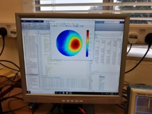
For simplicity, a great example of where UST is used can be found in ultrasound-based mammograms. When a tumour is present in the breast it affects the way in which ultrasound travels from one side of the breast to the other in two ways: changing the speed of the ultrasound or attenuating its amplitude. My system aimed to measure the change in velocity. When the difference in velocity is measured from many angles, an image of the breast along with any anomalous elements (tumours) can be created, and therefore the position of any tumours can be observed.
Getting started
This project was a very ambitious one as the university hadn’t constructed a system like this before. Unfortunately, the only commercial UST device at the university was tiny and sadly had been broken for years. So, the project had to start mostly from scratch. Thankfully, however, the department had many contacts from both Universities and industry that proved to be very useful. During my time on the project, I also had the privilege to meet many of these contacts when I attended the 9th World Congress on Industrial Process Tomography chaired by my supervising professor.
Attending this event really highlighted the power of partnership between institutions with much of the research being published at this global event having some form of collaboration. It could be anything from sharing test results from expensive equipment to having researchers visit for months to work with another university. For this project, collaboration with other groups of researchers was invaluable. A researcher now working in an industrial company based in Poland greatly accelerated the early development period as I didn’t have to start from complete scratch and could do away with much of the anticipated R&D.
The initial system
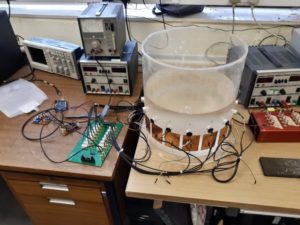
After a good while of tinkering and playing around with wires, I managed to start observing the transmission of ultrasound from one transducer to another through an acrylic tank filled with water. This initial system was driven by an Arduino driving a high-frequency audio amp that I made, and the receiving transducer’s waveform was observed using an oscilloscope. The system worked okay, but the amplifier did tend to overheat producing small amounts of smoke when really driven hard and burned me a good number of times.
Following this, I designed some dedicated boards to generate power amplified high frequencies, and then another board to amplify any received signals and tell the Arduino when a signal was received. As discussed before UST needs many readings from many angles, this in practice means there must be many transducers distributed around the observation area. I didn’t want to make a ton of my little boards, so instead, I designed a printed circuit board (PCB) that could choose which pair of transducers I wanted to use at a given point in time. This board was sent off to a fabrication plant and arrived 2 weeks later. Upon arrival of the PCB, I soldered everything up and placed 16 transducers around the same acrylic tank that I used earlier. Again, all processing in this instance is done with an Arduino that controls all the other boards.
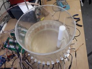
A limitation of this system was the degradation in image quality as the size of the inclusions in the tank decreased. This is attributed mainly to the number of transducers, and therefore the number of velocity samples taken from the tank. The only way to alleviate this issue is simple: increase the number of transducers. Which is exactly what we did and increased the total transducers from 16 to 32.
Developing the second system
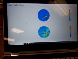
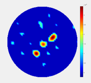
The true extent of the systems’ capabilities is currently being studied with some notable recent experiments showing that we can observe small crystals in the water along with temperature variations in the system. We also conducted some scans of biological tissue and constructed low-resolution images of bones in a human forearm, and fingers in a hand facing down the tank.
Academic publications
Due to the success of the system, several academic papers are being written regarding post-processing of its images and raw data, all of which I will be a co-author on, and one I am writing myself as lead author. My paper demonstrates the use of machine learning, specifically a convolutional neural network, to clarify and reconstruct images allowing isolation of individual objects. My paper proposes a method of generating training data for the neural network via simulation and then demonstrates that it can correct real UST images. This paper will be submitted for approval to Nature: Scientific Reports, hopefully in the coming weeks. Once this paper is concluded, myself and the other members of the project will investigate whether this technique is feasible for MRI scans and mammograms in isolating tumours or other anomalous biological objects.
Another notable paper being written, by a PhD student now back in China, is providing a method for combining UST and electrical-impedance-tomography (EIT) for the first time. The copper pads on the tank in previous images are used for EIT.
Thankfully this project has been successful and is providing many areas for additional research. It’s greatly improved many of my design skills and unquestionably helped me in my degree. I can’t recommend being involved in projects such as this enough, as it provides you with an opportunity to learn from experienced and gifted academics in a way that is not possible in a regular degree environment.
Respond